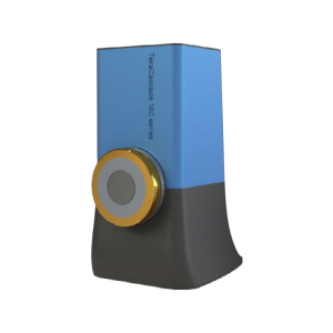
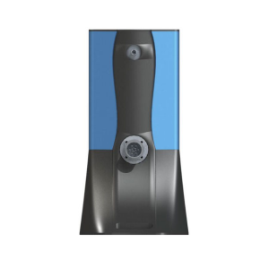
OcuScience iVivo VET-OCT Series OCT Imaging Systems
Function Overview :
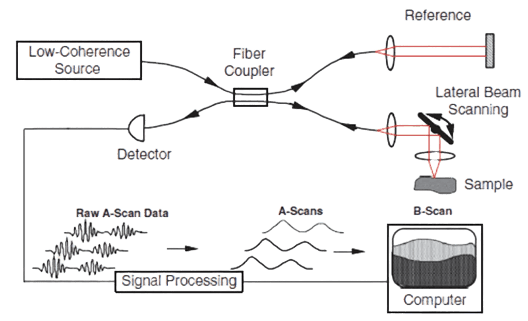
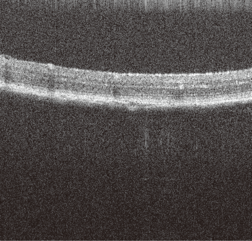
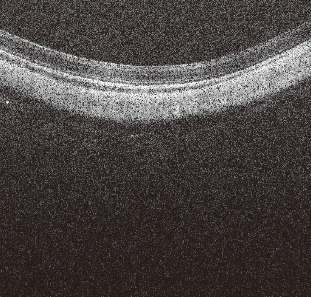
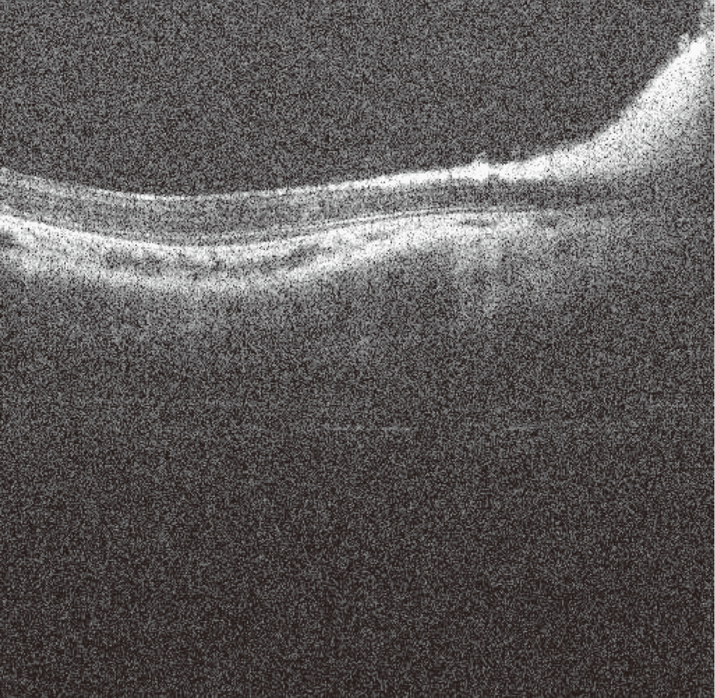
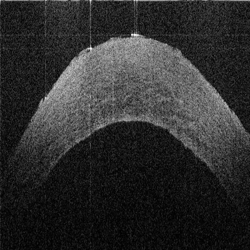
The OcuScience iVivo VET-OCT series is a high-precision imaging system designed for animal ophthalmology research and diagnostics, utilizing an infrared broadband light source and OCT technology to achieve micrometer resolution and millimeter depth of penetration, making it suitable for multi-species testing needs. The system combines a user-friendly interface with powerful software and multifunctional accessories to provide 3D tomography and fast rendering. With its flexible scanner and customizable modular design, the system is not only suitable for laboratories and veterinary clinics, but also supports surgical examinations and special studies, making it the best choice for animal ophthalmology research and diagnostics.
Description
Key Features
● Designed for small animal research: with interchangeable optics, a single system can be optimized for multiple species, including mice, dogs, horses, and rabbits, and is versatile enough to be used in laboratories, clinics, surgeries, and veterinary exams.
● Flexible and compact system: 3D printed housing, the whole system is installed in a blue metal plate shell, the shell is only the size of a shoebox, integrated micro PC computer (19*33*15 cm³, 2.7KG), is the multifunctional, smallest size of ophthalmology OCT products, flexible handheld scanner and touch screen console to make the animal examination easy!
● Powerful supporting software: The supporting software interface is user-friendly, intuitive, and easy to use, providing two measurement modes: real-time mouse scale analysis and automatic layer thickness tool, and integrating fast 3D scanning and rendering functions at the same time. It is also equipped with VQ Enhance, Intelligent Averaging and Alignment software to effectively improve image quality.
● Accessory package options: with endoscope, microscope, integrated viewing camera and other accessory packages.
● Customizable: workstation configuration - microscopic displacement stage, portable package configuration - handheld version, different light source wavelengths, scanning speeds
New energy lithium battery field
In the new energy industry, high-performance flexible pack battery electrode tape often has a multi-layer structure.
OCT has the ability to detect and recognize different layers of polymer material through penetration, reflecting the fit in real time.
Biotechnology and Life Sciences
Most of the internal details of biological tissues can be clearly imaged by infrared light-based OCT.
Therefore, OCT products are commonly used for cornea, eyeball, retina, skin, teeth and other research.
Manufacturing
Due to the high accuracy of optical interferometry, OCT is often used to evaluate the quality of agricultural products,
Measuring pill capsule characteristics, analyzing film coating thickness, and detecting cell phone folding screens,
Camera defects, fruit/drug detection, etc.
Integration Projects
Due to the compact and easy-to-integrate nature of the OCT optical path.
It can also be used for coaxial monitoring of laser processing and industrial/medical endoscopy.
Product Advantages
80KHz A scan rate
Fastest imaging animal ophthalmology OCT on the market
Versatile Design Non-Invasive Inspection
Suitable for laboratory, clinic, surgery, veterinary examination
Flexible for animal inspection
Equipped with a flexible handheld scanner and touch screen console
3μm axial resolution
More imaging details than other higher cost OCT systems
Open Platform and Information
Access to a wide range of powerful analytical tools
Smallest size ophthalmic OCT
The whole system is about the size of a shoebox.
Imaging of sample layers by Spectral Domain Optical Coherence Tomography, or SD-OCT.
Since its introduction in the 1990s, optical coherence tomography (OCT) has become an important tool for noninvasive tomography in the field of biology. Through low-coherence interferometry, OCT can generate high-resolution two-dimensional cross-sectional images from optically scattered signals from microstructures inside tissues in a manner similar to ultrasonic pulse echoes, achieving micrometer-level vertical and lateral resolution, and even capturing reflected signals as small as ~10^-10 optical power.
OCT excels in the detection of internal details of transparent, translucent and turbid materials, and it can also scan the surface contours of impenetrable materials, demonstrating a unique advantage in identifying surface and internal defects. Based on this advanced technology, OQ LabScope 3.0 SD-OCT is a compact system that produces high-resolution images of up to 512×512 pixels with a depth resolution of 2 microns. Its excellent price/performance ratio makes it comparable to OCT systems costing three times more, providing an ideal solution for researchers with limited budgets and helping them realize breakthrough research results.
Designed specifically for small animal research to achieve more accurate image presentation.
OCT technology has the advantages of non-contact, non-invasive, non-invasive, no damage/radiation, high imaging resolution, fast imaging speed, high sensitivity, good real-time performance, three-dimensional imaging, easy to combine with endoscopy technology, simple operation, etc. Since its invention nearly 30 years ago, this technology has been gradually expanded from the biomedical field to the nondestructive testing research, industrial production, as well as food and drug application areas.
OcuScience iVivo VET-OCT series is designed for this type of research, with an increased axial resolution of 3μm for more accurate image presentation. image presentation at scanning rates up to 80KHz.
Mouse Retina
Rabbit Retina
Rabbit Vision
Pig Cornea
Companion Software: Intuitive and Powerful Imaging and Analysis Tools
The companion software for the Honghong OCT Imaging System provides users with intuitive and powerful imaging and analysis tools designed to enhance the efficiency of your research and the quality of your results.
The software features an easy-to-use interface design that ensures high user-friendliness and is equipped with a variety of flexible scanning path modes, including landscape, portrait, cross and ring scanning, that users can freely choose from according to their needs.
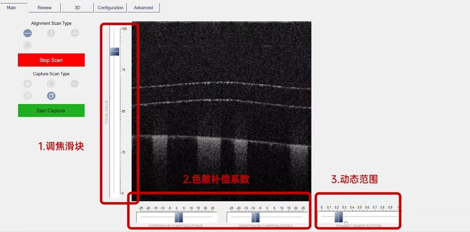
Flexible scanning path mode:
Supporting horizontal, vertical, cross and ring scanning, users can adjust the focus, dynamic range and other parameters according to specific needs to obtain the best image quality.
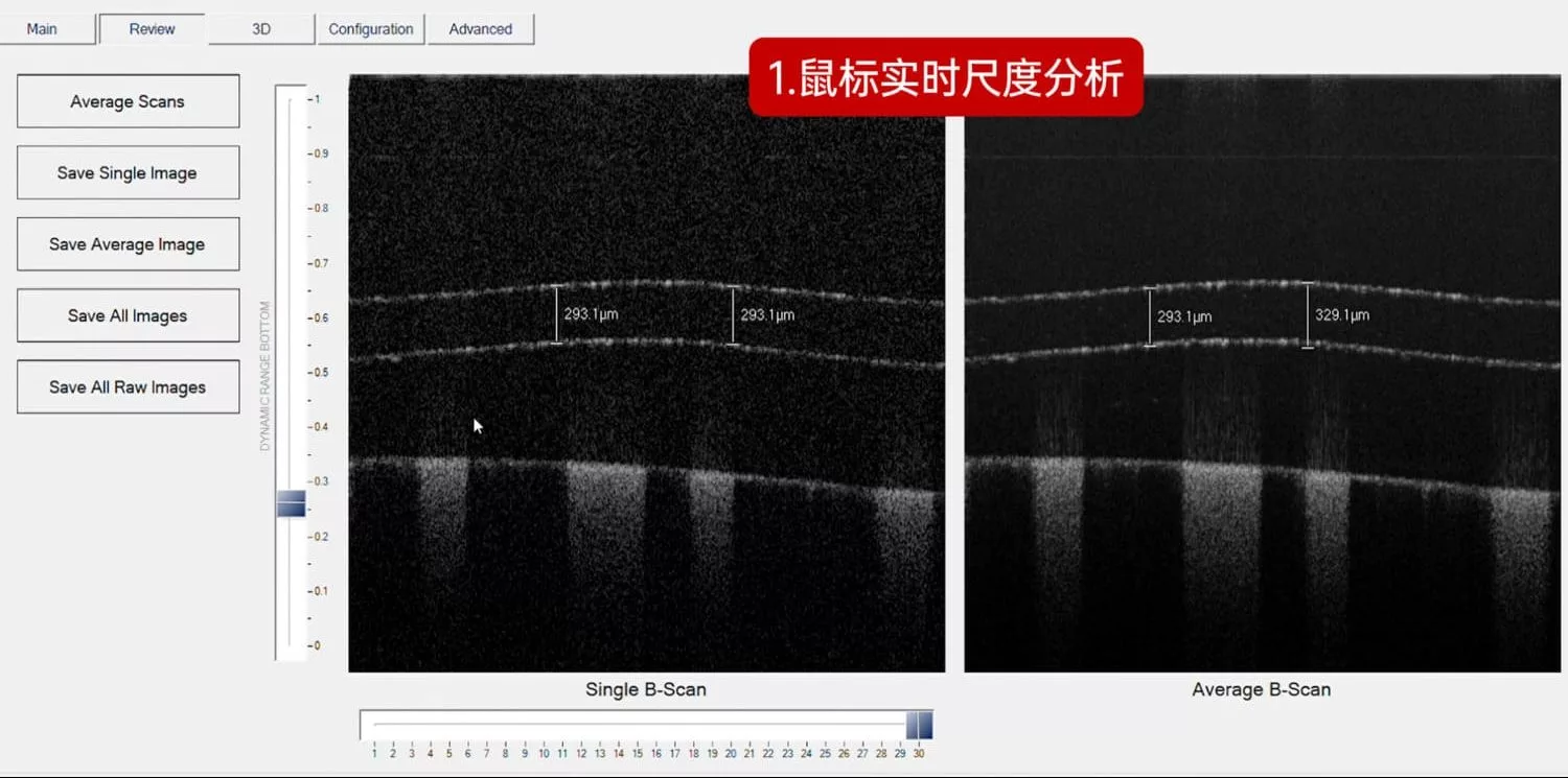
Immediate scale analysis:
With mouse operation, you can draw lines on the scanned image at any scale for precise detail measurements. This feature allows the user to quickly and accurately obtain the required data, greatly enhancing the flexibility of the analysis.
Automatic Layer Thickness Tool:
The software has a built-in automatic layer thickness analysis tool that automatically calculates the average thickness of a uniform cross-section and the thickness of segments, simplifying the complex thickness measurement process and providing users with accurate data support.
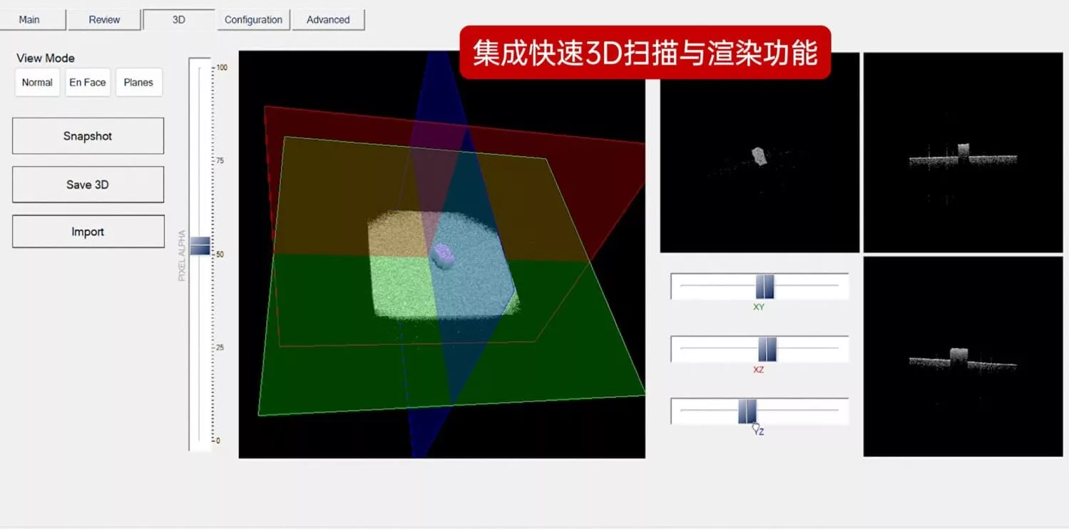
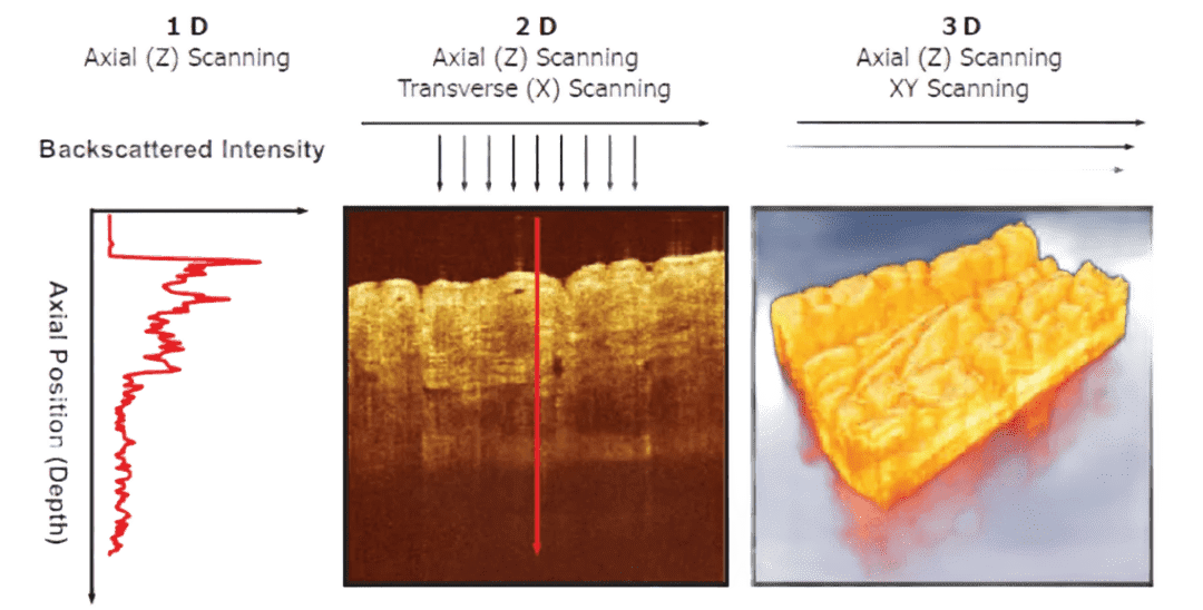
Fast 3D scanning and rendering:
Integrating efficient 3D scanning and rendering features, users can specify the scanning density and complete 3D reconstruction of up to 512 images in about 10 seconds. The software also supports drag-and-drop vector surfaces, allowing users to analyze any section in a 3D model, providing depth and all-around perspective.
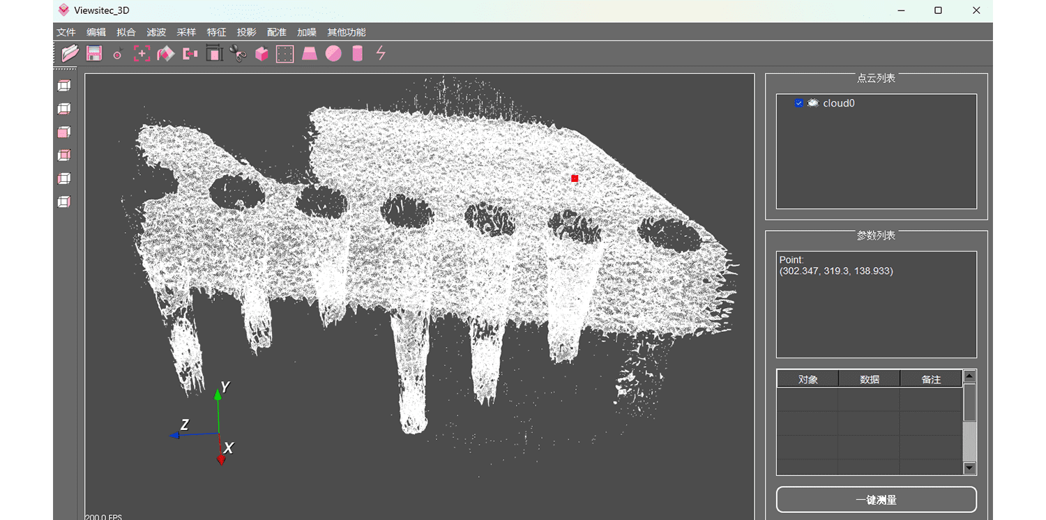
Row Selection Forms
| Model | 3.0 | X | R | XRD | SX | OQ StrataScope |
|---|---|---|---|---|---|---|
| performances | Base Model | higher speed | higher resolution | High speed + high resolution + deeper imaging | Ultra High Resolution + Higher Imaging Speed | Extreme depth imaging range in high scattering media |
| Center Wavelength | 840 nm | 840 nm | 860 nm | 860 nm | 890 nm | 1310 nm |
| Imaging Depth (Air/Organization) | 3.0 mm / 2.1 mm | 2.8 mm / 2.0 mm | 1.6 mm / 1.2 mm | 3.1 mm / 2.2 mm | 2.0 mm / 1.4 mm | 6.0 mm / 4.5 mm |
| Depth (axial) Resolution (in air/in tissue) | 8 / 6 μm | 8 / 6 μm | 5 / 3 μm | 5 / 3 μm | 3 / 2 μm | 14 / 10 μm |
| Horizontal Resolution | 18 μm | 18 μm | 18 μm | 18 μm | 18 μm | 20 μm |
| Line Scan Range | 7 mm | 7 mm | 7 mm | 7 mm | 7 mm | 8 mm |
| Scanning Scope | 5 x 5 mm² | 5 x 5 mm² | 5 x 5 mm² | 5 x 5 mm² | 5 x 5 mm² | 6 x 6 mm² |
| A-Scan | 34 kHz | 80 kHz | 34 kHz | 80 kHz | 80 kHz | 16 kHz |
| B-Scan frame rate | 22 fps | 50 fps | 22 fps | 50 fps | 50 fps | 11 fps |
Performance Upgrade Accessories
(1) 4× magnification objective: 5 mm line scan range, 3.5 mm x 3.5 mm body scan range
(2) 10× magnification objective: 2.5 mm line scan range, 1.8 mm x 1.8 mm body scan range
(3) 40× magnification objective: 1 mm line scan range, 0.7 mm×0.7 mm body scan range
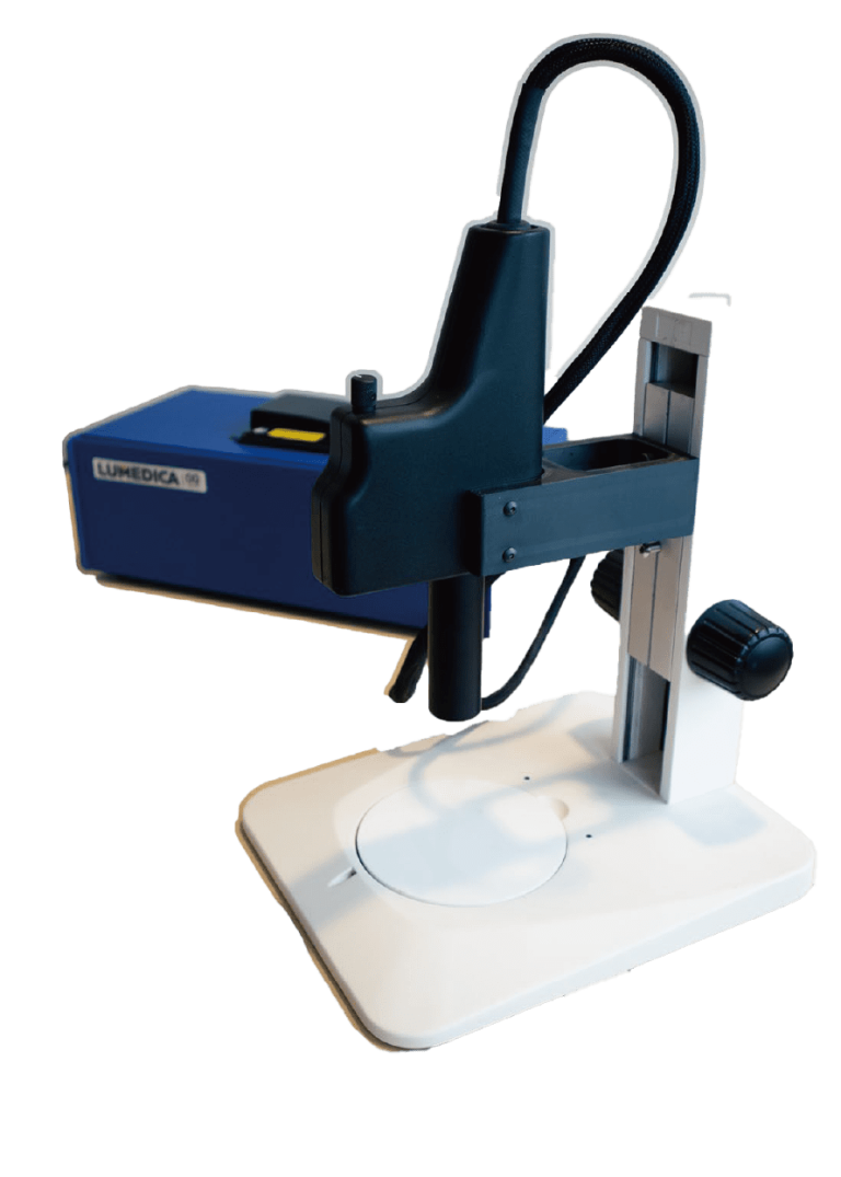
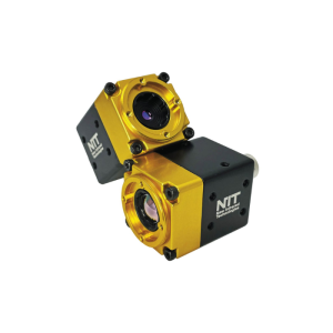
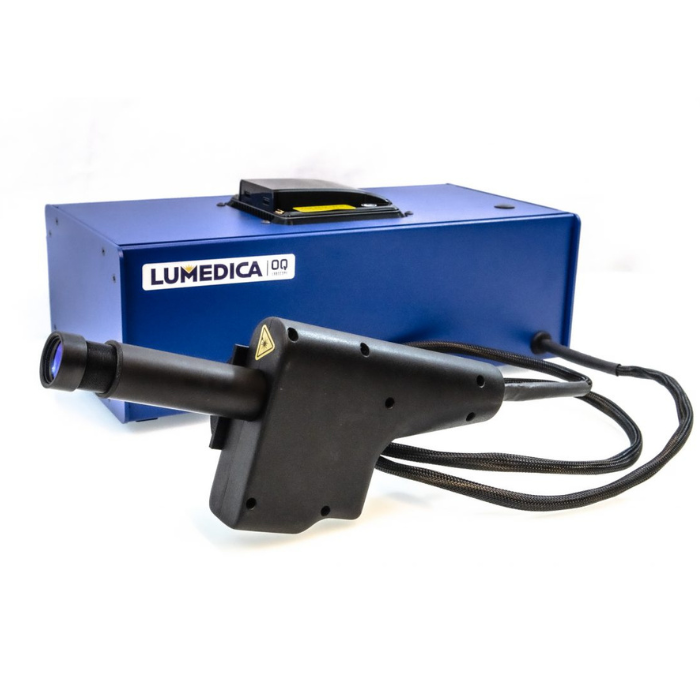
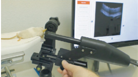
1.Microscope
Integration of a visible camera in the scanner for orientation and guidance

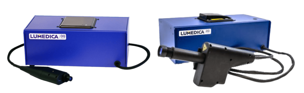
2. Standard Scanner & Scanner with integrated coaxial view camera
(1) Orthoscopic endoscope with a diameter of 4.2 mm and a field of view of 42 degrees.
(2) Side-viewing endoscope, 6 mm in diameter, with a scanning area of approximately 2 mm by 2 mm.

3.Customized Endoscope Scanning Heads

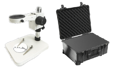
4.Sample table lifting and adjusting bracket, product safety box
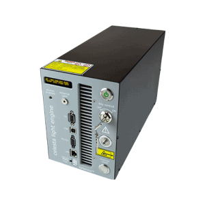
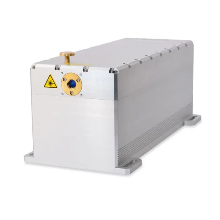
Upgraded 15 mW high power

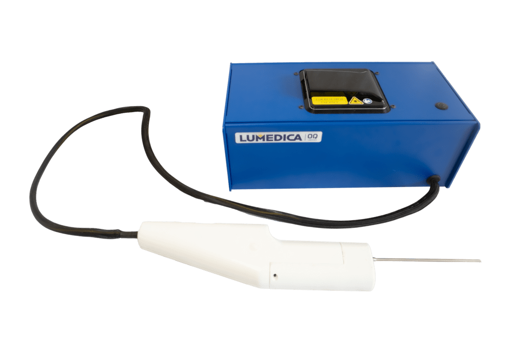
5.High luminosity light source

6.Optical Modules for Retinal Imaging
Reinforcement of swept cables with industrial-grade protective conduits

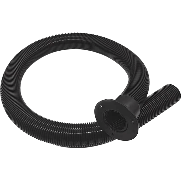
7.Industrial Grade Cables

8.Small Animal Holder

9.C#Source Code
Honghong will provide you with any support you need!
Our professional Honghong team will be the first to respond and provide you with the best service to solve all your problems.


